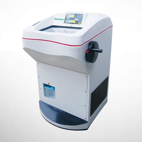
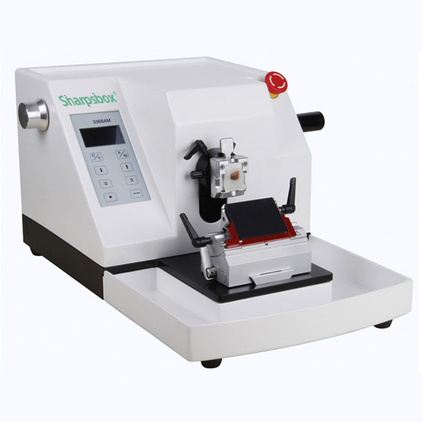
Touch Screen Cryostat Microtome
Cryostat Microtome features convenient operation and a modern fully automatic rotary microtome, 24 easily accessible freezing positions and well illuminated cooling chamber that can be effectively cooled down to -40 °C.
| Model: | DCM3000 | Capacity: | Max specimen size 55mm×80mm | Product Size: | 650×805×1160mm | Packing Qty: | 1unit | Carton Size: |
|---|
'SharpsBox' #DCM3000 model Cryostat Microtome features convenient operation and a modern fully automatic rotary microtome. For extra convenience the system includes a spacious stainless-steel chamber, 24 easily accessible freezing positions and well illuminated cooling chamber that can be effectively cooled down to -40 °C.
Features
■ This model has an ergonomic design and is manufactured using numerically-controlled machine tools;
■ A specimen retraction function protects the specimen from blade damage;
■ A trim button allows for easy switching between trim and sectioning mode;
■ Includes a counting function for the number of sections and the total thickness;
■ The cryogenic refrigeration system utilizes a forced cooling structure. The refrigeration chamber, freezing shelf, blade holder, and specimen holder are cooled independently by a German double compressor to enhance cooling performance and increase cooling speed.
■ Imported R404A non-CFC refrigerants thatare environment-friendly;
■ Achieves the desirable sectioning temperature within 20 minutes after turning on the unit when using the fast cooling mode;
■ UV option enables surface disinfection of harmful bacteria, viruses, and spores by using35 min after each use; It also has low-voltage DC shadowless lamp;
■ Semiconductor cooling function can be turned on and off;
■ Hearable and removable glass door;
■ Two defrosting modes: time defrosting and manual defrosting;
■ Microtome is outside of the refrigeration chamber to avoid the effect of heat-expansion and cold-contraction, thus minimizing the need for maintenance;
■ User-friendly intelligent operational interface is easy to learn and operate;
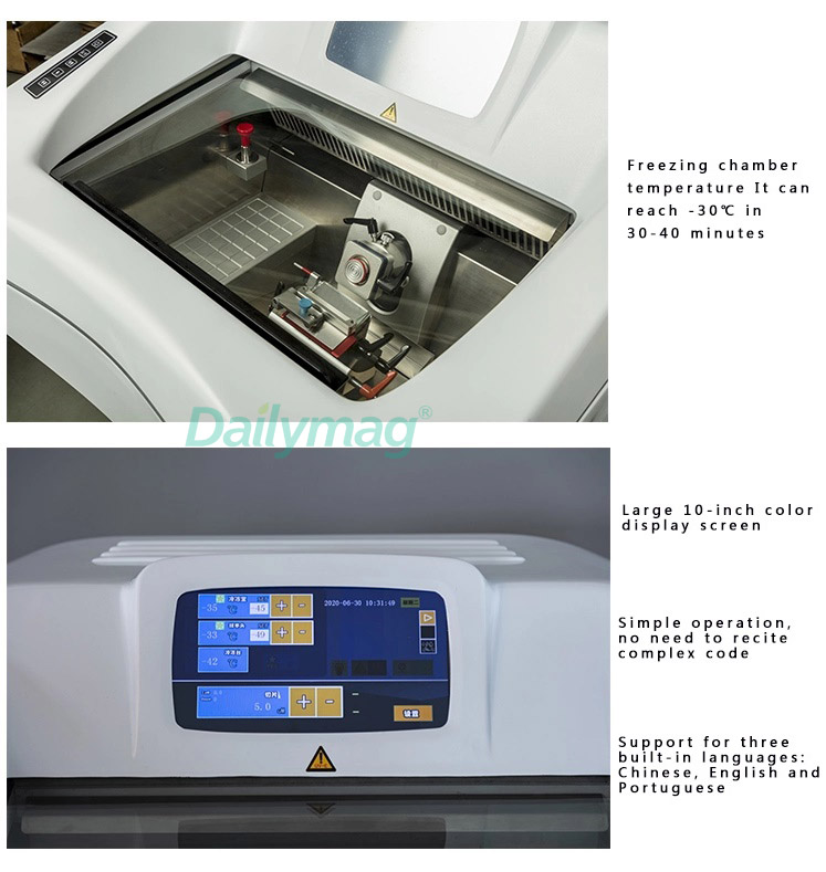
■ LCD screen displays the number of sections and the total thickness, section thickness, specimen retraction value, temperature setting, date, time, temperature and on/off timerare all included on the main display;
■ Auto sleep function results in longer compressor life: the cryo-chambertemperature automatically stays in a range of -1 to -9℃ under the sleep mode, and returns to the sectioning temperature within 15 minutes after canceling the sleep mode;
■ Keyboard locking function to avoid accident misoperation;
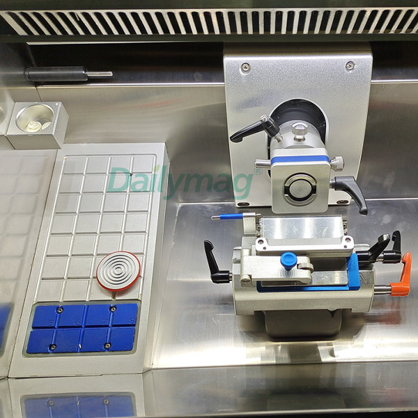
■ A mechanical hand-wheel lock ensure safe chamber access, 16 position for locking;
■ The red bar on the blade holder covers the whole length of blade to protect the user and the push bar enables easy changing of the blade;
■ A large-size quick-freezing shelf allows for the preparation of 26specimens at the same time; Two holes for semiconductor refrigeration;
■ It has cleaning device, easy help customer to clean the unit;
■ Specimen Orientation:XY – 12°; Z– 360° adjustable at any orientations to ensure precise placement;
■ Self-detection function for the temperature senor, can helpto warn the failure on temperature senor.

'SharpsBox' #DCM3000 model Cryostat Microtome Parameter
| Time to reach -40℃of the quick-freeze shelf: 60 minutes Lowest possible temperature of the Peltier element: -45℃ Lowest possible temperature of the Peltier elementtogether with quick-freeze shelf: below -60℃ Running time of the Peltier element:15minutes Maximum Specimen Size: 35mm×35mm Specimen Vertical Stroke: 60mm Specimen Horizontal Stroke: 20mm Electric Coarse Feed: rapid 7mm/s; slow 0.35mm/s |
| * The section thickness is adjusted in a range of 1μm to100μm * Trim Section Thickness Feed: adjusted in the range of 10μm to 400μm |
| * The section thickness is adjusted in a range of 1μm to100μm * Specimen Retraction Distance: adjustable in a range of 0 ~ 80μm, in 5μm increments * Working Voltage: AC220V±10%50Hz (standard model); AC110V±10%60Hz * Power: 600W * Dimensions: 650×805×1160mm(W×D×H) * Net Weight: 115kg |

What is Cryostat Microtome?
A cryostat refers to an instrument that can quickly make tissue slices for observation by making the tissue to a certain hardness at a low temperature. The cryostat consists of a slicing device and a refrigeration system. Usually, a complete cryosectioning system is composed of the main engine (freezing box, sample head, quick-freezing table, etc.), knife holder and blade; some cryostats also have a vacuum device, which is used to absorb waste inside the machine and use auxiliary equipment. The ability to flatten slices.
The principle and characteristics of Cryostat Microtome:
Cryostat is a technical method that uses physical cooling to freeze fresh tissue specimens to produce a certain hardness for slicing. Compared with paraffin sections, frozen sections do not need dehydration and embedding, so the preparation speed is fast, and it is a good method to provide pathological diagnosis for clinicians in operation. In addition, because cryosectioned specimens are fresh, unfixed tissue, cryostating is also an ideal preparation method for fat staining, enzymatic histochemical staining, and certain immunohistochemical staining and in situ molecular hybridization. The disadvantage of cryosection is that the morphology of tissue cells is slightly inferior to that of paraffin section.
Cryosection Operation Technology - How to Operate a Cryostat Microtome?
Frozen sections can be used for tissue sections such as fresh, formaldehyde-fixed, and cryo-refrigerated. The operation of frozen section generally goes through the steps of sampling, tissue fixation or non-fixation, quick freezing, frozen section, sticking or patch, staining and so on.
(1) Frozen sectioning
The tissues submitted for inspection should be as fresh as possible, and representative tissues should not contain too much water, so as to avoid tissue deformation caused by the formation of ice crystals; it should not be too large, too thick, and it is difficult to cut completely. 2mm is appropriate; generally not more than 3mm. Use a cryostat microtome to slice, and the tissue is preferably not fixed.
(2) Slicing step
The steps for slicing with a cryostat at constant temperature are as follows.
1. The cryostat should always be in the running state and cannot be turned on and off, so as not to affect the life of the machine. The temperature is generally adjusted to -18~-25°C (the temperature of the slicer can be set at -10°C at night). The freezing temperature of different tissues varies from -12 to -16°C for brain, lymph nodes, liver, spleen, kidney, etc.; -12--16°C for adrenal gland, thyroid, muscle, ovary, etc.; breast, skin, prostate, Ziguan etc. is -22~ -28°C. Insufficient freezing, the slices may be porridge-like or not cut out; excessive freezing, the slices will be crumbly or streaked.
2. Before slicing, put the tissue holder and the appropriate number of slicing knives on the freezing table for use.
3. The fresh tissue removed by surgery should not be fixed (because the fixed tissue is prone to produce large ice crystals, which is not conducive to slicing and affects staining), cut it into a thickness of 0.2 cm, and dry the blood and water on the surface of the tissue with a clean gauze.
4. Take out the frozen tissue tray, add a small amount of OCT (optimum cutting temperature) cryosection embedding medium or carboxymethyl fiber (or ordinary glue) on it, and put the taken tissue on it for quick freezing.
5. Put the tissue holder on the freezer table in the box and freeze for 1 to 2 minutes, then fix the tissue holder on the head of the microtome and adjust the position of the head so that it is just behind the slicer.
6. Roughly cut and trim the cut surface, then finely cut a few pieces of tissue on the knife with a brush, and adjust the fine-tuning scale to 5~6μm.
7. Adjust the anti-roll plate and start slicing. Carefully and accurately adjust the anti-roll plate to the proper position. When slicing, the cut slices can smoothly pass through the channel between the knife and the anti-roll plate and lie flat on the iron plate of the knife holder. At this time, the anti-roll plate can be lifted, and a glass slide can be attached to the section. Slides for attaching sections should be stored at room temperature, not in a freezer. Because when attaching slices, there is a temperature difference between the slides at room temperature and the slices in the freezer. When they touch each other, the adsorption force generated by the molecular transfer between them makes the slices and the slides firmly attached. paste.
8. The sections were fixed in methanol or AAF solution for 30-60s, washed with water, and then stained with HE.
9. After the frozen section is completed, the remaining tissue is removed and fixed for routine sectioning.
10. Clean up the tissue debris in the freezer, and turn on the ultraviolet lamp regularly for disinfection.
Related Products
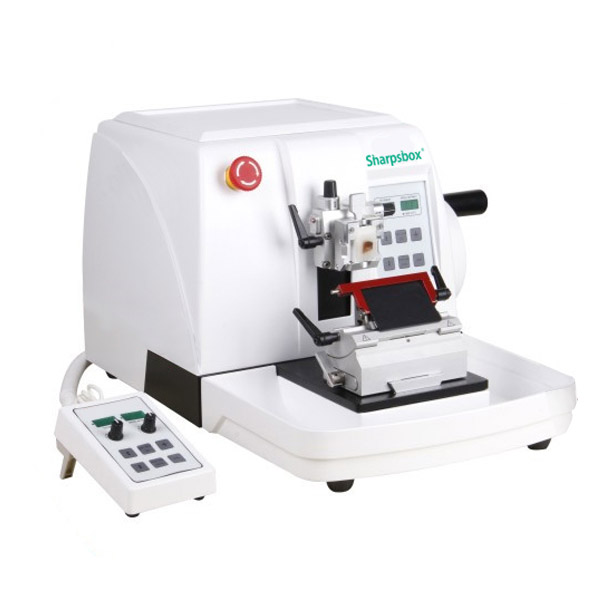
Premium Automated Rotary Microtome